Minimizing cariogenic organisms in the oral cavity is critical to the success of many dental procedures, and the maintenance of oral health.
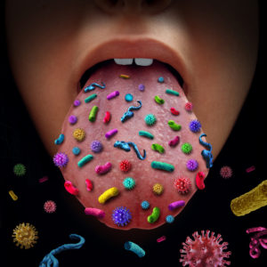
The oral cavity contains diverse microbiological communities. We have an estimated 20 billion microbes and over 1000 species in our mouths. Pathogenic bacteria and fungi found in the oral cavity can cause gum disease, bad breath, and periodontal problems standing in the way of your patient’s health.
Live microscopy can be a valuable tool in educating your patients about their own oral microbiome, assessing problems, and tracking improvements.
“Periodontitis is a disease associated with alterations in the subgingival microbiome. This significant oral disease affects approximately 47% of American adults and more than 740 million people in the world with an increasing prevalence as people age (6, 7).
“Studies of the subgingival microbial community identified a link between taxonomic composition and disease pathogenesis. The classic study by Socransky et al. revealed disease associations of specific bacterial organisms, including Porphyromonas, Treponema, and Tannerella, which were classified as the red complex organisms (13). Recent studies using 16S rRNA gene sequencing analysis provided a more comprehensive view of the subgingival community in health and disease. By analyzing samples pooled from different tooth sites and/or individuals, several case-control studies revealed distinct differences in the taxonomic composition for healthy and diseased states (14–17).” – Dynamic Changes in the Subgingival Microbiome and Their Potential for Diagnosis and Prognosis of Periodontitis[Internet]. Department of Molecular and Medical Pharmacology, Crump Institute for Molecular Imaging, David Geffen School of Medicine, UCLA, Los Angeles, California, USA. [cited 2018 Mar 01] Available from: http://mbio.asm.org/content/6/1/e01926-14.full
“While oral microorganisms exist in a symbiotic capacity, maintaining relationships with the host based on mutual benefits, some can transition to pathogens when they breach the barrier of commensalism, causing disruption of oral homeostasis, or ‘dysbiosis’ [3, 5]. Despite advances in our knowledge of the healthy oral microbiome, the functional aspects that lead to dysbiosis remain largely unknown [6]. What is now clear, however, is that oral diseases arise as a result of a change in the proportion of certain species with greater pathogenic potential within the indigenous flora [5]. This change in the ‘commensal’ microbiota is accompanied by disruption of the host immune homeostasis and development of an inflammatory response. Therefore, it is the prevalence of a certain combination of microbial species coupled with the inability of the host to contain their proliferation that is more indicative of a risk to develop disease.” – Sultan AS, Kong EF, Rizk AM, Jabra-Rizk MA (2018) The oral microbiome: A Lesson in coexistence. PLoS Pathog 14(1): e1006719. Available from: http://journals.plos.org/plospathogens/article?id=10.1371/journal.ppat.1006719
“Microbiological diagnosis of endogenous infections such as periodontal disease […] employ a system of microscopically counting bacterial morphotypes in wet mount preparations 2 and Gram-stained smears […] using cocci and increased motile rods and spirochaetes to differentiate between healthy and periodontally diseased sites in the mouth […].
“Spirochaetes are the predominant morphotypes microscopically observed in periodontal disease, 2,17 but because of their fastidious growth requirements, they are often not detected in cultural studies.
“Grade II morphotypes indicated increased spirochaetes and motile rods with an increase in PD from 1 to 2 mm demonstrating a transient phase, with a change in the ecology from health towards disease. Grade III morphotypes with an increase in spirochaetes and motile rods and a concomitant decrease in cocci and non-motile rods in sites with PD 3–4 mm could indicate an established gingivitis. Motile rods such as the capnophilic Gram-negative bacteria Capnocytophaga, Selenomonas and Wolinella are known to predominate in gingivitis lesions […].” – Kim Smeda-Pienaar, Eveline Kaambo & Charlene W J Africa (2017) Bacterial morphotype grading for periodontal disease assessment[Internet]. [cited 2018 Mar 01] Available from: https://www.nature.com/articles/bdjopen201611